


What Is a Slit Lamp Exam & Is It Important?
A slit lamp exam is a crucial diagnostic test that eye doctors use to check your eyes for problems.
This test can help detect eye conditions like cataracts and glaucoma before they worsen and cause vision loss.
You can treat issues quickly to prevent vision loss if you discover problems early.
Let’s explore the significance of the slit lamp exam in greater detail.
What is a slit lamp exam?
Eye doctors conduct a slit lamp exam to examine your eye’s different parts closely.
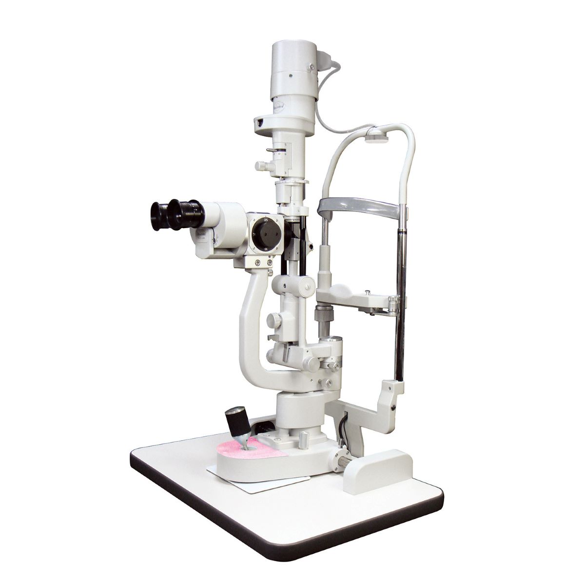
They use a unique slit lamp microscope, a specialized instrument combining a high-intensity light source and a magnifying lens.
It shines a narrow beam of light into your eye, allowing them to see things more clearly.
This exam helps doctors check the front part of your eye, like the cornea and the lens. They can also look at the back of your eye, like the retina and the optic nerve.
This test is essential because it can help doctors find any problems with your eyes before they worsen and cause vision problems.
How a slit lamp exam works

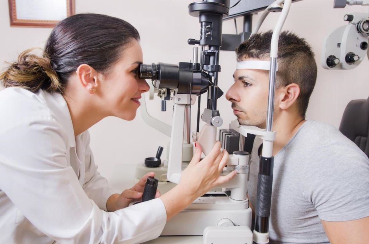
During a slit lamp exam, the eye doctor will have you sit in front of the machine with your chin resting on support and your forehead against a bar.
They will then shine a narrow beam of light onto your eye and use the slit lamp microscope to look at the different parts of your eye.
The front part of your eye, like the cornea and the lens, is examined for abnormalities. These abnormalities include scratches, infections, or cataracts.
The eye doctor will also check the back part of your eye, like the retina and the optic nerve.
Compared to other eye examination methods, such as a regular eye exam or a retinal exam, the slit lamp exam provides a more detailed view of the eye’s structures.
The results can help the eye doctor diagnose and monitor different eye conditions more accurately. It’s also non-invasive and painless.
Why slit lamp exams are necessary
Supporting cataract surgery
The slit lamp exam can also support eye surgeries like cataract surgery. Before the surgery, your eye doctor will perform a thorough slit lamp exam to get an in-depth look at the structures of your eyes.
This same diagnostic technique will be used throughout recovery to ensure everything is healing correctly.
Posterior segment condition diagnosis
The slit lamp exam is beneficial for examining the eye’s posterior segment, including the retina, optic nerve, and vitreous.
This part of the eye is difficult to see with a regular eye exam and requires a more detailed examination.
Possible diagnosis with a slit lamp exam
A slit lamp exam is a standard procedure in ophthalmology to diagnose and monitor various eye conditions.
Here are some of the possible diagnoses from a slit lamp exam:
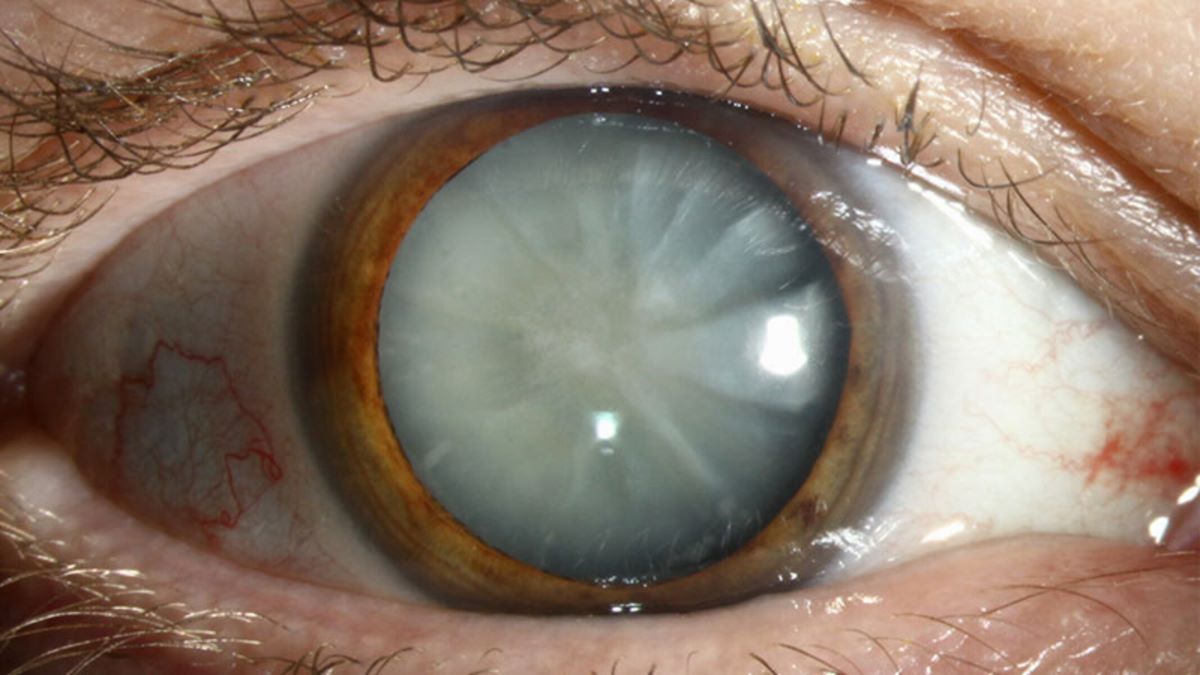
Cataracts
The clouding of the lens, which characterizes cataracts, can be visualized in detail during a slit lamp exam.
Glaucoma
A slit lamp exam is essential to glaucoma diagnosis and monitoring, as it helps detect glaucoma by allowing the doctor to observe the optic nerve head and retinal nerve fiber layer.
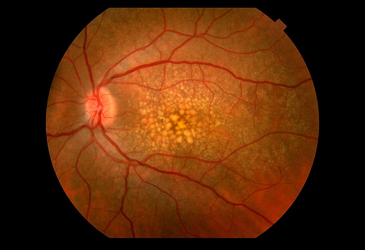
Macular degeneration
A slit lamp exam can also aid in diagnosing macular degeneration by allowing doctors to examine the macula.
That’s part of the eye that is responsible for central vision.
Diabetic retinopathy
Diabetic retinopathy is a condition that can occur as a complication of diabetes and can cause damage to the blood vessels found in the retina. When left untreated, it can result in vision loss.
A slit lamp exam can help detect diabetic retinopathy by revealing changes or abnormalities in the retina’s blood vessels.

Corneal ulcers
A slit lamp exam can help diagnose corneal ulcers by revealing the location and size of the ulcer.
Conjunctivitis
Conjunctivitis, or pink eye, can be diagnosed using a slit lamp exam. During this exam, an eye doctor can identify signs of inflammation in the conjunctiva.
Some potential symptoms include redness, swelling, or discharge.
Corneal abnormalities
A slit lamp exam can help diagnose corneal abnormalities such as scars, infections, or dystrophies by providing detailed visualization of cornea layers.
In short – slit lamp exams are essential for eye health
A slit lamp exam is a valuable diagnostic tool that allows eye doctors to examine the eye’s structures in detail and detect various eye conditions.
It can help preserve your vision and improve your eye health by detecting and treating these conditions early.
Keep your eyes healthy and functioning by scheduling regular eye exams — including a slit lamp exam.
Early detection of potential conditions can help you preserve clarity and longevity in sight.
References
- “Slit Lamp Exam”, Healthline.
- “Uses of the Slit Lamp Parts of the Slit Lamp”. University of Illinois in Chicago.
- “Using the slitlamp to perform cataract surgery on upright patients”. Journal of Cataract and refractive surgery.
- “Recognising and managing diabetic retinopathy”. National Library of Medicine.
- “Keratitis”. Mayo Clinic.
- “Slit Lamp Microscopy”. Science Direct.

Written by:
Angie Garcia